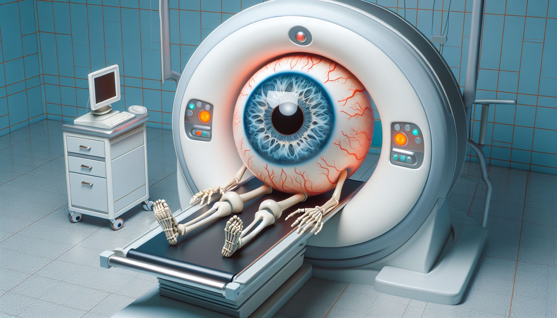
Age-Related Macular Degeneration (AMD) is a leading cause of vision loss among the elderly, with its more severe form, Wet AMD, being particularly aggressive. Wet AMD occurs when abnormal blood vessels grow under the retina and macula, leading to rapid and severe vision loss if untreated. The advent of Optical Coherence Tomography (OCT) has revolutionized the management of Wet AMD, providing a non-invasive, detailed view of the retina that is critical for both diagnosis and treatment monitoring.
Diagnosis with OCT
OCT uses light waves to take cross-section pictures of the retina, allowing ophthalmologists to see the layers of the retina and measure their thickness. These images are crucial in diagnosing Wet AMD, as they can reveal the presence of fluid or blood beneath the retina—hallmarks of the disease. OCT imaging can also detect the buildup of drusen, tiny lipid or protein deposits, which are early indicators of AMD.
Here’s what patients typically experience during an OCT procedure:
- Non-Invasiveness: Patients find comfort in the fact that OCT is non-invasive. Unlike some other eye tests, there are no needles, dyes, or physical contact with the eye.
- Quick and Painless: The procedure is relatively quick, lasting only a few minutes per eye. Patients sit in front of the OCT machine, and a technician captures detailed cross-sectional images of the retina using low-intensity light. There is no pain or discomfort during the scan.
- Fixation Target: Patients are asked to focus on a central point (usually a small light) to ensure accurate imaging. This helps stabilize eye movement during the scan.
- No Need for Dilation: Unlike some retinal exams that require pupil dilation, OCT imaging can often be done without dilation. This is a relief for patients who dislike the temporary blurriness caused by dilating eye drops.
- Quiet Environment: The room where the OCT is performed is usually quiet and dimly lit. Patients may hear soft clicks as the machine captures images, but it’s generally a calm and serene environment.
- Results and Discussion: After the scan, patients receive immediate results. The technician or ophthalmologist explains the findings, discusses any abnormalities, and outlines the next steps if necessary.
- Routine Monitoring: For patients with conditions like Wet AMD, regular OCT scans are essential for monitoring disease progression. Patients become accustomed to these follow-up visits and appreciate the proactive approach to managing their eye health.
The cost of OCT imaging:
- Cost of OCT Imaging:
- The cost of OCT imaging can vary significantly based on factors such as the type of device, features, and brand. High-end OCT systems with advanced capabilities tend to be more expensive.
- Traditional tabletop OCT devices used in ophthalmology clinics can range from several thousand to tens of thousands of dollars.
- However, there are also low-cost and portable OCT systems available. For example, biomedical engineers have developed a portable OCT scanner that aims to bring this technology to underserved regions 1. These systems are more affordable and accessible.
- Accessibility to Patients:
- The accessibility of OCT imaging to patients depends on several factors:
- Clinical Setting: In well-established eye care centers, OCT imaging is readily accessible. However, smaller clinics or remote areas may have limited access.
- Specialty and Scope of Practice: Optometrists and ophthalmologists use OCT differently. Some optometrists use it primarily as a screening tool, referring patients to specialists when abnormalities are detected. Others diagnose and manage a broader range of diseases.
- Advancements in Technology: As technology evolves, more affordable and portable OCT devices are becoming available. These innovations enhance accessibility.
- Geographic Location: In regions with advanced healthcare infrastructure, patients have better access to OCT. Conversely, underserved areas may face challenges.
- Research and Development: Ongoing efforts aim to make OCT more accessible globally, especially in resource-limited settings.
- The accessibility of OCT imaging to patients depends on several factors:
- Advancements in Accessibility:
- Researchers have developed low-cost, portable OCT scanners that extend beyond ophthalmology. These systems can be used in hard-to-reach areas, including joints and other body parts 2.
- The goal is to democratize OCT technology, making it available to a broader patient population.
Treatment Monitoring
Once Wet AMD is diagnosed, treatment typically involves anti-VEGF therapy, which helps reduce the growth of abnormal blood vessels and slow the leakage that causes retinal damage. OCT is indispensable in monitoring the retina’s response to these treatments. By regularly imaging the retina with OCT, doctors can assess whether the medication is effectively reducing fluid accumulation and can adjust treatment plans accordingly.
Research and Future Directions
OCT is not only a tool for current clinical practice but also a window into the future of Wet AMD management. Ongoing research is leveraging OCT to better understand the pathophysiology of Wet AMD and to develop new treatments. With advancements in OCT technology, such as OCT angiography, doctors can now visualize blood flow in the retina without the need for dye injections, further improving the safety and comfort of patient care.
Conclusion
The integration of OCT into the management of Wet AMD represents a significant advancement in ophthalmology. It provides a safe, accurate, and detailed method for diagnosing Wet AMD and monitoring its treatment. As technology progresses, OCT will undoubtedly continue to play a vital role in preserving vision and improving the quality of life for patients with Wet AMD.
Here are more sources that discuss the use of Optical Coherence Tomography (OCT) in diagnosing and managing wet Age-Related Macular Degeneration (AMD):
- Mayo Clinic provides details on how OCT is used in the diagnosis and treatment of wet macular degeneration, including monitoring retina response to treatments1.
- amdbook.org offers an in-depth look at OCT technology in Age-related Macular Degeneration, explaining how it allows for detailed analysis of anatomic structures and neovascular membrane lesions subtypes2.
- WebMD explains how doctors use OCT to diagnose wet AMD, including checking for changes in the retina that can be signs of AMD and deciding if the retina will respond to treatment3.


